Research
Different imaging, quantifying and analytical methods and their specific preparation procedures are needed to work on the research topics of the division Evolution and Biogeography of Meiofauna that are introduced on the further subpages.
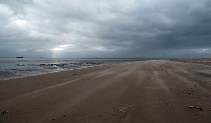
At the beginning of any research, however, we need to procure samples with suitable objects for our studies. In the simplest case, this might be a sand sample from a beach or a plankton catch from a weeded pond, which can be obtained with relatively little effort. If sediment of deeper water depths is needed, the sampling can be done with snorkelling or diving equipment, or a grab has to be used from a boat. If sediment from oceanic biotopes has to be examined (e.g. deep sea floor, shelf around offshore islands, summit plateaus of seamounts), the effort is considerably greater. In such a case, sampling devices such as a usual Van Veen grab up to remotely operated vehicles (ROVs) need to be deployed from large research ships.
The sediment samples with the containing organisms are chemically fixed immediately. However, fragile taxa such as the gastrotrichs mostly have to be extracted from the sediment alive in order to be prepared carefully for the respective examination method. Different microscopic procedures are available for morphological investigations. These range from conventional light microscopy, fluorescence and confocal laser scanning microscopy to high-resolution scanning and transmission electron microscopy. The analysis of DNA sequences of different genes is meanwhile an important tool for phylogeographic, taxonomic and systematic problems. A reliable identification of various species in an environmental sample via specific gene fragments is becoming more and more important in an age of rapidly advancing, man-made environmental change.